Biomedical Laboratory Science

ShareThis

Tuesday, January 23, 2018

Cutaneous Leishmaniasis: Immune Responses in Protection and Pathogenesis.


Cutaneous leishmaniasis is a major public health problem and causes a range of diseases from self-healing infections to chronic disfiguring disease. Currently, there is no vaccine for leishmaniasis, and drug therapy is often ineffective. Since the discovery of CD4+ T helper 1 (TH1) cells and TH2 cells 30 years ago, studies of cutaneous leishmaniasis in mice have answered basic immunological questions concerning the development and maintenance of CD4+ T cell subsets. However, new strategies for controlling the human disease have not been forthcoming. Nevertheless, advances in our knowledge of the cells that participate in protection against Leishmania infection and the cells that mediate increased pathology have highlighted new approaches for vaccine development and immunotherapy. In this Review, we discuss the early events associated with infection, the CD4+ T cells that mediate protective immunity and the pathological role that CD8+ T cells can have in cutaneous leishmaniasis.


Cutaneous leishmaniasis — which is caused by several protozoal parasites of the genus Leishmania — is endemic to South and Central America, Northern Africa, the Middle East and parts of Asia, and an estimated 1 million new cases arise each year. Of particular interest to immunologists is the wide range of clinical manifestations associated with this disease, which, similar to tuberculosis and leprosy, is dictated largely by the type and magnitude of the immune response of the host. As in most infections, the immune response to cutaneous leishmaniasis depends on many host factors, as well as on the differences between the infecting Leishmania spp. Experimental infections in mice also exhibit a spectrum of clinical presentations depending on the mouse strain and the infecting parasite species or strain used.
  • Cutaneous leishmaniasis exhibits a wide spectrum of clinical presentations that is determined largely by the host immune response. The host immune response to infection is influenced both by host genetics and the Leishmania spp. and/or strain.
  • The rapid recruitment of neutrophils and inflammatory monocytes following infection with Leishmania influences the course of disease. Neutrophils can have both protective and deleterious roles, whereas inflammatory monocytes kill Leishmania parasites and differentiate into monocyte-derived dendritic cells that promote the development of protective CD4+ T helper 1 (TH1) cells.
  • Control of Leishmania infection depends on the production of interferon-γ by CD4+ TH1 cells, which leads to enhanced killing by macrophages due to the production of reactive oxygen species and nitric oxide.
  • CD8+ T cells recruited to Leishmania lesions exhibit a cytolytic profile and lyse infected cells without killing the parasites, which leads to enhanced inflammation and increased severity of disease. Controlling these pathogenic CD8+ T cells, or the downstream mediators of inflammation that they induce, is a new approach to leishmaniasis immunotherapy.
  • Infection with Leishmania generates several types of CD4+ T cells that mediate resistance to reinfection, including effector T cells, effector memory T cells, central memory T cells and tissue-resident memory T cells. There is currently no Leishmania vaccine, and a hurdle for vaccine development is that the most effective T cells are short-lived effector T cells; targeting longer lived central memory and tissue-resident memory T cells is an alternative approach.

Monday, January 22, 2018

Kaplan MCAT Biology Review - Test Prep !

MCAT Biology Review,

Test Prep for the MCAT Exam

KAPLAN pdf


The MCAT is changing in 2015. With the addition of three semesters’ worth of material, more advanced critical thinking skills, a longer duration, and changes in Biology content, the new exam requires even more diligent prep with resources from Kaplan Test Prep.

Kaplan's MCAT Biology Review offers:


Sunday, January 14, 2018

Cancer Transcriptome Profiling at the Juncture of Clinical Translation !


Methodological breakthroughs over the past four decades have repeatedly revolutionized transcriptome profiling. Using RNA sequencing (RNA-seq), it has now become possible to sequence and quantify the transcriptional outputs of individual cells or thousands of samples. These transcriptomes provide a link between cellular phenotypes and their molecular underpinnings, such as mutations. In the context of cancer, this link represents an opportunity to dissect the complexity and heterogeneity of tumours and to discover new biomarkers or therapeutic strategies. Here, we review the rationale, methodology and translational impact of transcriptome profiling in cancer.


Transcriptomics is the large-scale study of RNA molecules by use of high-throughput techniques. It examines the abundance and makeup of a cell's transcriptome. In contrast to DNA, which is largely identical across all cells of an organism, the actively transcribed RNA is highly dynamic, reflecting the diversity of cell types, cellular states and regulatory mechanisms. Because a transcriptome profile can be regarded as a signature or snapshot of the underlying cell state, the experimental profiling of samples and specimens can provide insights into their unique biology.

Depending on the specific approach, transcriptomics can not only reveal the architecture of gene expression but also provide details on the structure, modification and variation of individual transcripts. Advances in transcriptome profiling, specifically the development of genome-wide methodologies targeting diverse RNA species, have enabled us to discover the seemingly endless complexity of RNA biology and to comprehensively annotate the human genome and other eukaryotic genomes. Arguably, transcriptomics is currently the most well-established modality and foundation of functional genomics, a field of study for which the goal is to synthesize large-scale data to understand the mechanisms that govern cellular and organismal phenotypes.
  • RNA sequencing (RNA-seq) has the potential to bridge tumour genotypes (for example, mutations) and their phenotypic consequences (for example, cancer molecular subtypes).
  • The field of transcriptomics has matured thanks to lockstep developments in experimental protocols, algorithms and databases.
  • Methodological and algorithmic advances continue to enable clinical applications of transcriptome profiling.
  • Detection of gene fusions is the most immediate application of RNA-seq.
  • Gene expression signatures have demonstrated prognostic and predictive value.
  • Transcriptome profiling will be essential for immuno-oncology.


Thursday, December 7, 2017

MCQ 12. 11 years old boy, restless all time, unable to concentrate in class, hardly ever in his seat, roams around the hall, difficulty in playing quietly.


MCQ 12. An 11 years old boy is all the time so restless that the rest of the class is unable to concentrate. He is hardly ever in his seat and roams around the hall. He has difficulty in playing quietly.


What is the the most likely diagnosis?
a. Attention-deficit hyperactivity disorder 
b. Conduct disorder 
c. Depressive disorder 
d. Schizophrenia


Correct answer: Click here

Wednesday, December 6, 2017

Transcriptional Regulation by Mediator Complex


Alterations in the regulation of gene expression are frequently associated with developmental diseases or cancer. Transcription activation is a key phenomenon in the regulation of gene expression.

In all eukaryotes, mediator of RNA polymerase II transcription (Mediator), a large complex with modular organization, is generally required for transcription by RNA polymerase II, and it regulates various steps of this process. The main function of Mediator is to transduce signals from the transcription activators bound to enhancer regions to the transcription machinery, which is assembled at promoters as the preinitiation complex (PIC) to control transcription initiation.


Recent functional studies of Mediator with the use of structural biology approaches and functional genomics have revealed new insights into Mediator activity and its regulation during transcription initiation, including how Mediator is recruited to transcription regulatory regions and how it interacts and cooperates with PIC components to assist in PIC assembly.

Novel roles of Mediator in the control of gene expression have also been revealed by showing its connection to the nuclear pore and linking Mediator to the regulation of gene positioning in the nuclear space. Clear links between Mediator subunits and disease have also encouraged studies to explore targeting of this complex as a potential therapeutic approach in cancer and fungal infections.


Key points

  • Recent structural advances based on improvements in electron microscopy methodology have enabled the generation of high-resolution structural models of the mediator of RNA polymerase II transcription (Mediator) complex and of the preinitiation complex (PIC) in the presence of Mediator.
  • The module composition of Mediator changes between its recruitment to upstream regulatory regions (enhancers or upstream activating sequences where Mediator is bound to transcription factors) and its action on core promoters together with PIC components.
  • The functional interplay between Mediator and general transcription factors in PIC assembly is closely related to chromatin architecture at promoter regions.
  • Direct contact between Mediator and the nuclear pore-associated transcription-coupled export (TREX2) complex suggests that Mediator functions in gene positioning in the nuclear space.
  • Mediator has been shown to function in the establishment of transcriptional memory, which also involves Mediator interactions with the nuclear pore.
  • Potential therapeutic targeting and modulation of Mediator activity in cancers and in fungal infectious diseases emphasizes the importance of studies of Mediator mechanisms for improving human health.


Saturday, December 2, 2017

Human Microbiome and its Association With Health and Diseases

Human microbiota are distinct communities of microorganisms that resides at different body niches. Exploration of the human microbiome has become a reality due to the availability of powerful metagenomics and metatranscriptomic analysis technologies. Recent advances in sequencing and bioinformatics over the past decade help provide a deep insight into the nature of the host-microbial interactions and identification of potential deriver genes and pathways associated with human health, well-being, and predisposition to different diseases. In the present review, we outline recent studies devoted to elucidate the possible link between the microbiota and various type of diseases. The present review also highlights the potential utilization of microbiota as a potential therapeutic option to treat a wide array of human diseases.

Humans are viewed as composites of human and microbial cells. Human microbiota are complex and dynamic microbial communities composed mainly of bacteria, but also includes protozoa, archaea, viruses, and fungi that resides in and on different body niches such as oral cavity, throat, esophagus, stomach, colon, urogenital tract, respiratory tract, and skin. The number of microbial cells inhabiting human body is estimated to exceed the H.sapiens cells by 10-fold and estimated at 350 trillion microbial cells.


Microbiome-host interactions. Schematic representation showing the perplexed microbial-host
interactions due to different triggering factors on microbiota and their genetic material constitute;
the human microbiome.
Source: Wiley

Friday, December 1, 2017

Herpes gladiatorum: Facts, Symptoms, Causes, Risk Factors, Diagnosis, Treatments, Prevention and Outlook.


Herpes gladiatorum is an infection caused by herpes simplex virus type 1, a virus similar to that which causes cold sores. It is common among those who engage in high-contact sports, such as wrestling, and is also known as mat herpes. Those infected with herpes will carry the virus with them for life as there is no cure.


There will be periods where the virus is inactive and no symptoms are present, and times where it is active and causes various symptoms and flare-ups.

When the virus is active, the person carrying it is infectious and can spread herpes gladiatorum to others. The virus can be transmitted by skin-to-skin contact and is easily passed from one person to the next.

In this article, we examine the symptoms of herpes gladiatorum and the parts of the body that can be affected. We also take a look at diagnosis, treatment, and prevention of the condition.


Thursday, November 30, 2017

How to Apply Today for Sanima Equity Fund Using Online ASBA Service (Step-by-Step Process of Global IME Bank Online ASBA)

SEBON had compulsorily implemented Applications Supported by Blocked Amount (ASBA) system from Shrawan 1, 2074 for public issues (IPO/FPO/MF) and Sanima Capital Ltd. is the next company issuing its Sanima Equity Fund from full ASBA services all over Nepal starting from Tuesday November 28th 2017. Global IME Bank (GBIME), like several other banks, has come up with online ASBA service registration of application for share with the aim to provide easy and efficient services to its customers.


ShareUpdateNepal


The customers can use ASBA services from either mobile banking or internet banking service without being presented at the bank and also at zero cost for the applications of IPO, FPO and Mutual Funds, such as, this Sanima Equity Fund.


Applicants having account in Global IME Bank can apply Sanima Equity Fund through online service for registration of application.


Here is a step-by-step process to apply for the Sanima Equity Fund using the online ASBA system:

STEP 1: Open Global IME Internet Banking by clicking on the link below.


STEP 2: Position the cursor at ASBA ONLINE at top right corner as shown.


STEP 3: Click login after entering your online banking username and password as shown.


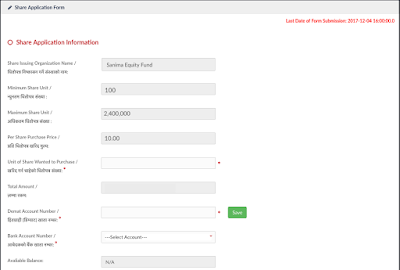
STEP 4: You will have 'Share Application Form' to enter your information details as shown as shown.


List of available public offerings along with details on the apply button of
the 
public offering for application submission.

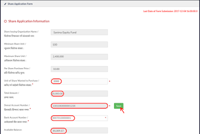
STEP 5: Complete the upper portion of the form with your information into the fields as shown.

Note at the bottom the 'available balance' of your account !


STEP 6:
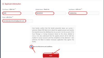
 Scroll down and complete the bottom portion of the form with your information into the fields as shown.


Read the terms and conditions in the box at the bottom,check the circle to mark
your agreement and thenclick 'Apply' in the red box at the bottom.


STEP 7: Click 'Confirm' (or 'Edit' if incorrect) for share conformation as shown.


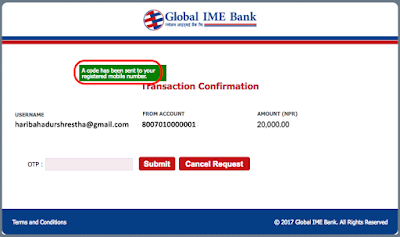
STEP 8: You will get a dialog box from the Global IME Bank on your screen displaying that 'A code has been sent to your registered mobile number' in green box with details as shown.


STEP 9: Enter into the 'OTP' box the 6 digit code sent to your registered mobile number and hit 'Submit' in the red box at the bottom as shown.


STEP 10: If everything goes well, you will get a 'Customer Verification' dialog box displaying 'Your account details has been verified successfully.', then hit the 'Submit Form' in the red box at the bottom as shown.


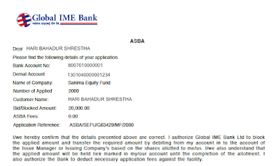
STEP 11: You will then be offered 'ASBA Receipt' dialog box with the 'PDF' icon link, click it to download your receipt as shown.


STEP 12: Finally, save the pdf ASBA receipt for your future reference as shown.


That is it! You are done!! Hope this helps & Good Luck!!!


ShareUpdateNepal: Thursday November 30th 2017


ALSO,  CHECK THESE OUT TOO!

Sanima Capital Issues Sanima Equity Fund from Nov 28 to Dec 1, 2017 !

Support Microfinance IPO Starting from Tomorrow, How is the Company?


Related Posts Plugin for WordPress, Blogger...

AddToAny