Biomedical Laboratory Science

ShareThis

Showing posts with label Gout. Show all posts

Tuesday, April 5, 2016

Atherosclerosis, Rheumatoid Arthritis and Inflammation.

Rheumatoid arthritis (RA) has long been associated with increased cardiovascular risk, but despite substantial improvements in disease management, mortality remains high. Atherosclerosis is more prevalent in RA than in the general population, and atherosclerotic lesions progress at a faster rate and might be more prone to rupture, causing clinical events. Cells and cytokines implicated in RA pathogenesis are also involved in the development and progression of atherosclerosis, which is generally recognized as an inflammatory condition. The two diseases also share genetic and environmental risk factors, which suggests that patients who develop RA might also be predisposed to developing cardiovascular disease. In RA, inflammation and atherosclerosis are closely linked. Inflammation mediates its effects on atherosclerosis both through modulation of traditional risk factors and by directly affecting the vessel wall. Treatments such as TNF inhibitors might have a beneficial effect on cardiovascular risk. However, whether this benefit is attributable to effective control of inflammation or whether targeting specific cytokines, implicated in atherosclerosis, provides additional risk reduction is unclear. Further knowledge of the predictors of cardiovascular risk, the effects of early control of inflammation and of drug-specific effects are likely to improve the recognition and management of cardiovascular risk in patients with RA.

Rheumatoid arthritis (RA) is associated with a significantly increased risk of cardiovascular mortality, accounted for mainly by increased atherosclerotic disease.1, 2 Although the prevalence of some traditional cardiovascular risk factors is increased in RA, adjustment for these factors does not fully account for the heightened risk, suggesting that RA itself is an independent risk factor for cardiovascular disease (CVD).3 The prevalence of atherosclerosis is increased in RA, even in early disease,4 and chronic inflammation is thought to promote atherosclerosis both by modulation of traditional risk factors and also possibly by direct biological effects on the artery. In this article, we discuss the potential mechanisms that might accelerate atherosclerosis in RA, with a particular focus on inflammation.

Read more: Atherosclerosis, Rheumatoid Arthritis and Inflammation.

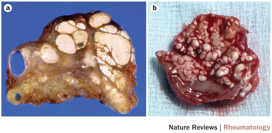
Figure 2: Development of an atherosclerotic plaque.
Source: NatureReviewsRheumatology

Global epidemiology of gout: prevalence, incidence and risk factors.

Gout is a crystal-deposition disease that results from chronic elevation of uric acid levels above the saturation point for monosodium urate (MSU) crystal formation. Initial presentation is mainly severely painful episodes of peripheral joint synovitis (acute self-limiting 'attacks') but joint damage and deformity, chronic usage-related pain and subcutaneous tophus deposition can eventually develop. The global burden of gout is substantial and seems to be increasing in many parts of the world over the past 50 years. However, methodological differences impair the comparison of gout epidemiology between countries. In this comprehensive Review, data from epidemiological studies from diverse regions of the world are synthesized to depict the geographic variation in gout prevalence and incidence. Key advances in the understanding of factors associated with increased risk of gout are also summarized. The collected data indicate that the distribution of gout is uneven across the globe, with prevalence being highest in Pacific countries. Developed countries tend to have a higher burden of gout than developing countries, and seem to have increasing prevalence and incidence of the disease. Some ethnic groups are particularly susceptible to gout, supporting the importance of genetic predisposition. Socioeconomic and dietary factors, as well as comorbidities and medications that can influence uric acid levels and/or facilitate MSU crystal formation, are also important in determining the risk of developing clinically evident gout.

Gout is the most common form of inflammatory arthritis and is caused by chronic elevation of serum uric acid (SUA) levels above the saturation point for monosodium urate (MSU) crystal formation. The deposition of MSU crystals, which occurs predominantly in peripheral joints and surrounding tissues, defines gout. The characteristic presentation is of rapidly developing monoarticular synovitis in peripheral joints (an acute 'attack') that is extremely painful but self-limiting, with resolution within several days or 1–2 weeks. However, long-term deposition of MSU crystals can result in joint damage and disfiguring subcutaneous tophi. In addition, gout is also associated with many conditions that affect longevity and well-being,1 such as metabolic syndrome,2 cardiovascular diseases3, 4, 5, 6 and renal diseases. In particular, gout is increasingly recognised as an independent cardiovascular risk factor.

Read more: Global epidemiology of gout: prevalence, incidence and risk factors.

Figure 1: The estimated prevalence of gout across the world.
Source: NatureReviewsRheumatology

A structural basis to stone formation in gout.

The mechanisms and sites of monosodium urate monohydrate (MSU) crystal deposition in gout have received little attention from the scientific community to date. Formalin fixation of tissues leads to the dissolution of MSU crystals, resulting in their absence from routinely processed pathological samples and hence neglect. However, modern imaging techniques—especially ultrasonography but also conventional CT and dual-energy CT—reveal that MSU crystals form at the cartilage surface as well as inside tendons and ligaments, often at insertion sites. Tophi comprise round white formations of different sizes surrounded by inflammatory tissue. Studies of fibres recovered from gouty synovial fluid indicate that these fibres are likely to be a primary site of crystal formation by templated nucleation, with crystals deposited parallel to the fibres forming transverse bands. In tophi, two areas can be distinguished: one where crystals are formed on cellular tissues and another consisting predominantly of crystals, where secondary nucleation seems to take place; this organization could explain how tophi can grow rapidly. From these observations based on a crystallographic approach, it seems that initial templated nucleation on structural fibres—probably collagen—followed at some sites by secondary nucleation could explain MSU crystal deposition in gout.

Gout is characterized by deposits of monosodium urate monohydrate (MSU) crystals, a consequence of hyperuricaemia—serum uric acid levels raised above normal and sodium urate reaching a concentration above supersaturation. Usually, crystals are formed in joints and periarticular tissues, the magnitude of the deposit growing and extending to other sites whilst hyperuricaemia persists. The mechanisms of pathological formation of MSU crystals on tissue have received scant attention to date. In this Review, we take a correlative structural approach to explore possible mechanisms of MSU crystal formation in gout on the basis of morphological findings.

Read more: A structural basis to stone formation in gout.

Figure 3: Formations of MSU crystals in tophi.
Source: NatureReviewsRheumatology
Related Posts Plugin for WordPress, Blogger...

AddToAny