
The mechanisms and sites of monosodium urate monohydrate (MSU) crystal deposition in gout have received little attention from the scientific community to date. Formalin fixation of tissues leads to the dissolution of MSU crystals, resulting in their absence from routinely processed pathological samples and hence neglect. However, modern imaging techniques—especially ultrasonography but also conventional CT and dual-energy CT—reveal that MSU crystals form at the cartilage surface as well as inside tendons and ligaments, often at insertion sites. Tophi comprise round white formations of different sizes surrounded by inflammatory tissue. Studies of fibres recovered from gouty synovial fluid indicate that these fibres are likely to be a primary site of crystal formation by templated nucleation, with crystals deposited parallel to the fibres forming transverse bands. In tophi, two areas can be distinguished: one where crystals are formed on cellular tissues and another consisting predominantly of crystals, where secondary nucleation seems to take place; this organization could explain how tophi can grow rapidly. From these observations based on a crystallographic approach, it seems that initial templated nucleation on structural fibres—probably collagen—followed at some sites by secondary nucleation could explain MSU crystal deposition in gout.
Gout is characterized by deposits of monosodium urate monohydrate (MSU) crystals, a consequence of hyperuricaemia—serum uric acid levels raised above normal and sodium urate reaching a concentration above supersaturation. Usually, crystals are formed in joints and periarticular tissues, the magnitude of the deposit growing and extending to other sites whilst hyperuricaemia persists. The mechanisms of pathological formation of MSU crystals on tissue have received scant attention to date. In this Review, we take a correlative structural approach to explore possible mechanisms of MSU crystal formation in gout on the basis of morphological findings.
Read more: A structural basis to stone formation in gout.
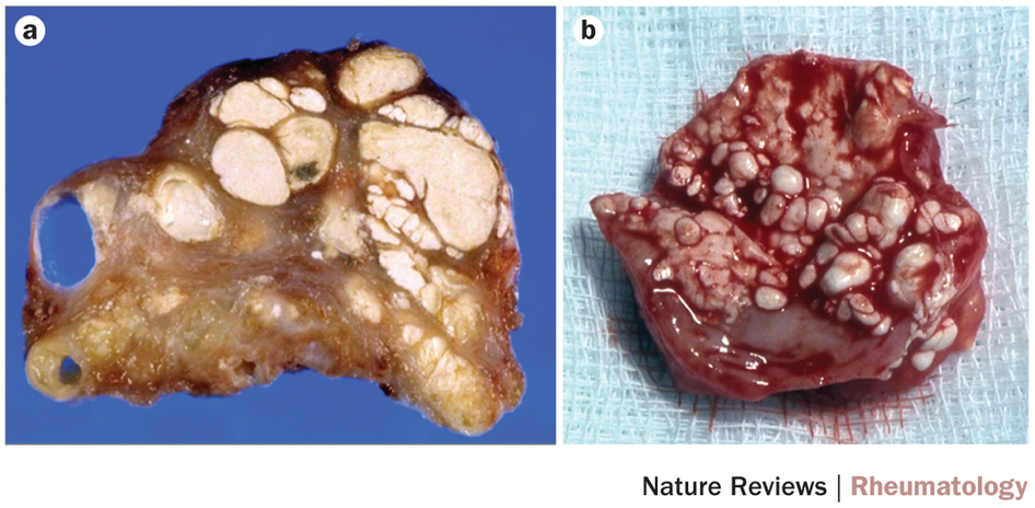 |
| Figure 3: Formations of MSU crystals in tophi. |
